File:Jmol Hin tetrad DNA.gif
From 2006.igem.org
No higher resolution available.
Jmol_Hin_tetrad_DNA.gif (550 × 550 pixels, file size: 42 KB, MIME type: image/gif)
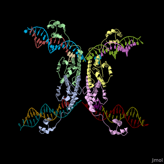
Protein Data Bank ID 1ZR4, Jmol image. A Hin protein dimer binds and cleaves DNA at each HixC sequence flanking the fragment of DNA to be inverted. The two dimers (dimer 1 = leftward green and blue protein structures; dimer 2 = rightward yellow and purple protein structures) come together to form a tetrad complex where DNA ends are swapped and ligated.
Li, W., Kamtekar, S., Xiong, Y., Sarkis, G.J., Grindley, N.D., Steitz, T.A. (2005) Structure of a synaptic gamma delta resolvase tetramer covalently linked to two cleaved DNAs. Science. v309 pp.1210-1215 , 2005
File history
Click on a date/time to view the file as it appeared at that time.
| Date/Time | Thumbnail | Dimensions | User | Comment | |
|---|---|---|---|---|---|
| current | 17:56, 26 October 2006 | | 550×550 (42 KB) | Kahaynes (Talk | contribs) | (Protein Data Bank ID 1ZR4, Jmol image. A Hin protein dimer binds and cleaves DNA at each HixC sequence flanking the fragment of DNA to be inverted. The two dimers (dimer 1 = leftward green and blue protein structures; dimer 2 = rightward yellow and purple) |
File links
The following page links to this file: